Bulletin News
02/05/2013
Aphrodite Ahmadi sees the laws of physics demonstrated in some unusual places, including in a baby frog’s eye.
An assistant professor of physics at SUNY Cortland, she recently examined microscopic slides of sectioned tadpole eyes to study the mechanics of a human genetic weakness that leads to a rare form of blindness.
Retinitis pigmentosa is as mysterious in its development as it is devastating in its effect on the person who slowly loses his or her sight from it at a relatively young age. It can erase vision from the center of a person’s line of sight to the periphery, from the periphery to the inside, or in an irregular pattern.
Presently, this form of blindness has no cure.
Ahmadi and colleagues at Upstate Medical University’s Neuroscience and Physiology Department and its Ophthalmology Department recently described clues about the disease’s progression.
The findings by Ahmadi and her co-researchers — Mohammad Haeri and Barry Knox — were published Jan. 22 in an article in Biophysical Journal titled “Modeling the Flexural Rigidity of Rod Photoreceptors.”
“Light-sensing cells in the eye rely on their outer segment to convert light into neural signals that allow us to see,” said Ahmadi, the study’s senior author, giving a physicist’s perspective. “But because of its unique cylindrical shape, the outer segment is prone to breakage, which can cause blindness in humans.”
The team’s new experimental and theoretical findings help to explain the mechanical properties that cause the outer segment to snap under pressure. The researchers are convinced that their findings point to the origin of some severe eye diseases and could lead to new ways of preventing blindness.
 |
| Aphrodite Ahmadi |
“To our knowledge, this is the first theory that explains how the structural rigidity of the outer segment can make it prone to damage,” Ahmadi said. “Our theory represents a significant advance in our understanding of retinal degenerative diseases.”
The outer segment of photoreceptors consists of hundreds of discs packed with a light-sensitive protein called rhodopsin, she explained. The discs produced by the body at nighttime are different from those produced during the day, generating a banding pattern that was first observed in frogs but is common across species. Mutations that affect photoreceptors often destabilize the outer segment and may damage its discs, leading to cell death, retinal degeneration, and blindness in humans.
“But until now, it was unclear which structural properties of the outer segment determine its susceptibility to damage,” Ahmadi said.
To address this question, Ahmadi and her team examined tadpole photoreceptors under the microscope while subjecting them to fluid forces. They found that high-density bands packed with a high concentration of rhodopsin were very rigid, which made them more susceptible to breakage than low-density bands consisting of less rhodopsin. Their model confirmed their experimental results and revealed factors that determine the critical force needed to break the outer segment.
 |
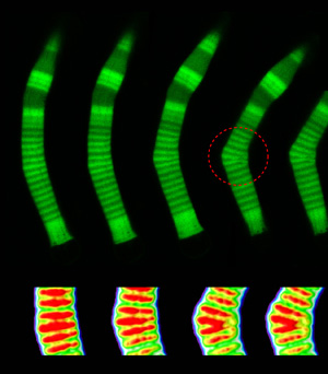
| SUNY Cortland Assistant Professor of Physics Aphrodite Ahmadi's study encompassed the basics of eye rod flexibility. This genetically fragile rod is shown bending as it begins to fail. The above left image, which details the rods bending, appeared in the Biophysical Journal and is reproduced here with the journal's permission. |
When looked at under a microscope, the rod photoreceptor cells in a tadpole eye appeared to gradually bend into a crook like a boomerang and break. Eventually, the rods no longer will transfer visual information to the brain.
“The findings support the idea that mutations causing rhodopsin to aggregate can destabilize the outer segment, eventually causing blindness,” she said. “Further refinement of the model could lead to novel ways to stabilize the outer segment and could delay the onset of blindness.”
Although her knowledge is central to the study, she started the collaboration almost by accident. Three years ago, soon after joining SUNY Cortland with a recent doctorate in physics from Syracuse University, she helped a fellow graduate student by proofreading his thesis. That collaborator was Haeri, currently a post-doctoral fellow in Upstate Medical University’s Neuroscience and Physiology Department.
“I was correcting spelling and such when I found out about these rods and photoreceptors,” Ahmadi said. “He was studying something different than this study: the biochemistry part about the gene mutation that leads to blindness. I looked at the rod structure and saw that some of them looked bent.
“As a physicist, I became interested in this phenomenon and started looking for a mechanical explanation,” she said.
Their research associate, Knox, is a professor and chair of neuroscience and physiology at Upstate.
“Haeri got approval from Dr. Knox to gather more images and for me to study the images — almost 300 of them — to figure out how they could bend and come up with a physical explanation for it.
“It was an interesting structure for me. The rods are supposed to be straight. Why should they break? They are like straightened fingers, with no place to jiggle around.”
Ahmadi has developed a model for the phenomenon she has observed but still is unclear on what added force generally breaks down retinas prone to this disability. However, she thinks that both internal and external forces could cause the eye structure’s collapse.
“As far as we know, there are some forces in the eye chamber,” she said. “Anything that can apply force can break it down. Any pressure to the eye can become a force.”
They chose to study Xenopus frogs that make one of a wide variety of genetic anomalies leading to this particular disease.
"We did this study to see how this bending and breakage depends on the concentration of rhodopsin along the cell,” she said. “In regions with higher concentration of rhodopsin the rod becomes less flexible and will break under pressure.”
In true physicist fashion, the paper features graphs to measure the rate of rod bending — or deflection — over time, depending on the level of rhodopsin density.
“Some graphs compare high density versus low density bends,” Ahmadi said. “The normal cell recovers its structure, but in a high density band it keeps bending and bending because it has lost its internal structure.”
Ahmadi’s study expenses were offset by $3,000 in SUNY Cortland faculty research grant funding, while the National Institutes of Health supported her colleagues for their laboratory work.
Ahmadi hopes to move closer to finding the origin of the forces responsible for the breakage of the rod cells in further investigations.
“For now, the model sounds pretty good and the results agree perfectly with what we see,” she said. “In the next step, we want to apply controlled forces to the rods and find the experimental value needed to break the rods. The magnitude of the forces can give us hints on what their origin might be.”
A cure is not on the immediate horizon, but at least humans are beginning to understand the disorder.
“This is actually the first study that I’m aware of that has been done on the mechanical cause of this blindness,” she said. “There is still a long way to go.”